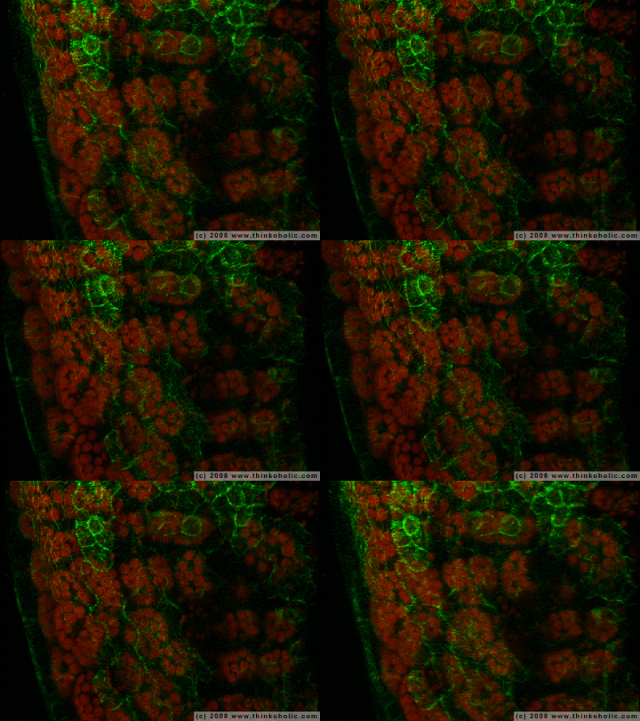
last week, i took part in a practical class dealing with specific kinds of microscopy – namely confocal laser scanning microscopy (CLSM) and transmission electron microscopy (TEM).
while we’re still waiting for the results of the second part, we’ve had plenty of time to play around with the CLSM.
here’s one of the numerous animations:
it shows part of a leaf of arabidopsis thaliana, that was genetically modified in a way that makes certain structure molecules within the cell walls visible.
to be more specific, the sequences of the green fluorescent protein (GFP, originally a gene of the crystal jellyfish, aequorea victoria) and a corresponding microtubule binding domain were inserted into the plant’s DNA, so that the cell walls would emit green light when exposed to light of a particular frequency (not all cells actually produced these molecules).
autofluorescense of chloroplasts is displayed in red.
using the CLSM, about 20 images were recorded at different levels of the leaf. these layers were then rendered in an animation that highlights its tridimensionality.
the image width equals 150 μm, that’s approx. 1/7th of a millimeter.

Very, very cool. Gratz :)